- Also known as the beta pleated sheet due to the pleated appearance of the protein structure from a side view.
- No strict rules to how they are formed because the hydrogen bonds can be formed between distant amide hydrogen and carbonyl oxygen.
- They are two or more strands distant from each other in the primary structure that form hydrogen bonds with each other side by side.
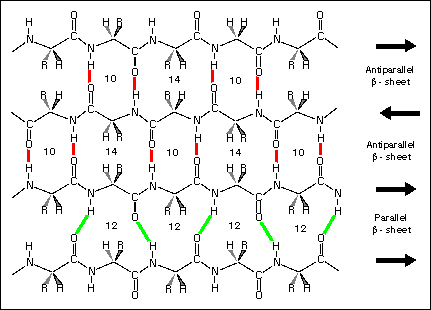
- There are two types of beta sheet structures; parallel and anti-parallel. Parallel beta sheets have strands that run in the same direction as each other and anti-parallel beta sheets have strands that run in opposite direction to each other. The hydrogen bonding in anti-parallel beta sheets are usually more linear.
- The N-H and C=O groups on the outer edge of the beta sheet structure are not hydrogen bonded to other strands of the primary sequence.
- If the R-groups along the outer edge of the beta sheets are polar, it can interact with solvents such as water. If they are non-polar, they can interact with hydrophobic structures such as lipids.
- They can also pack closely against side chains of nearby alpha helix structures.
- Almost all of the polar amide groups are hydrogen bonded to each other in the beta sheet structure.
- Parallel sheets are almost always buried in the inside of the structure of a protein and anti-parallel sheets are mostly exposed to solvent due to the amino acids that make up that part of the structure. Therefore, anti-parallel sheets are seen as being more stable structures than parallel sheets.
- Parallel sheets usually have other structures, such as helices, separating them from other parallel sheets.
- They have a right handed twist to the beta strands due to the steric factors of the L-amino acid configuration.
- Isoleucine and valine are often found in these beta sheets because they are hydrophobic.
- Beta strands can be amphipathic because of the alternating side chains of amino acids next to each other. These amphipathic strands are found on the surface of proteins.
- A large anti-parallel beta sheet can also form a barrel structures (such as retinol binding protein). The last strand of the beta sheet is hydrogen bonded to the first strand so it forms a closed barrel shape.
- The exterior of the structure is usually surrounded by solvent as it is hydrophilic and the interior is where the hydrophobic residues are found so non-polar species can be found here (e.g retinol).
Monday, 5 November 2012
Secondary Structure of Proteins (Beta sheets)
Beta sheets
Subscribe to:
Post Comments (Atom)
no left handed in beta sheet?
ReplyDelete